|
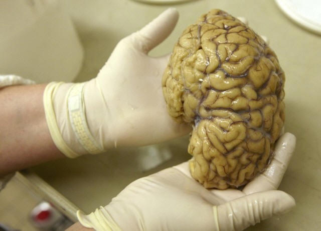
 Researchers examined results from what’s known as positron emission
tomography (PET) scans for four current and 10 former National
Football League (NFL) players who had at least one previous
concussion as well as for 16 similar men who weren’t athletes and
had no history of concussions. Researchers examined results from what’s known as positron emission
tomography (PET) scans for four current and 10 former National
Football League (NFL) players who had at least one previous
concussion as well as for 16 similar men who weren’t athletes and
had no history of concussions.
They measured levels of a substance called translocator protein
18KDa (TSPO), which are thought to rise when the brain responds to
traumatic injuries.
Compared with men who weren’t in the NFL, the football players had
higher levels of TSPO and greater changes in the brain’s white
matter, the study found.
“The study showed that there is a measurable degree of this
biomarker of brain injury and repair even in young NFL players,
suggesting that the insult to their brains could have occurred long
before they were scanned for the study – perhaps dating to
collegiate or pre-collegiate play,” said senior study author Dr.
Martin Pomper, a researcher at Johns Hopkins Medical School in
Baltimore.

“That could be a cautionary note for young athletes participating in
contact sports,” Pomper added by email.
While more research is needed to confirm the results from this small
study, one day it may be possible to use PET to look for TSPO and
determine which athletes might be at risk for neurological or
psychiatric problems after a head trauma, Pomper said.
One of the most vexing issues with treating concussions in athletes
is that the full extent of brain injuries can be difficult to assess
while players are still alive. In particular, a condition tied to
sports concussions known as chronic traumatic encephalopathy (CTE)
can only be diagnosed during an autopsy.
For the current study, researchers focused on resident immune cells
of the central nervous system called microglia that are thought to
play a role in the brain's response to injury and other
neurodegenerative processes.
Scientists think it’s possible prolonged microglial activation can
happen after single or and repeated traumatic brain injuries. When
this happens, normally low levels of TSPO in brain tissue may rise.
[to top of second column] |

The NFL players, who reported an average of seven years since their
last concussion, showed higher TSPO in 8 of 12 regions of the brain
examined in the study, researchers report in JAMA Neurology.
Beyond its small size, other limitations of the study include a lack
of data to explain exactly when TSPO levels rose relative to the
timing of brain injuries, the authors note.
Even so, the results point to a potential way to better pinpoint the
extent of brain damage in living athletes, as well as the
possibility of one day of developing experimental treatments to
target inflammation in the brain as tool for minimizing the health
impact of concussions, Jonathan Godbout of Ohio State Wexner Medical
Center in Columbus writes in an accompanying editorial.
“These findings are not limited to NFL players,” Godbout said by
email. “Prolonged brain inflammation (chronic microglia activation)
is likely a key component to myriad neurological diseases and
perhaps even normal brain aging.”
“Like anything, if you can identify the source of the problem prior
to the development of neurological complications then you can
intervene,” Godbout added. “How to intervene in humans and target
this inflammatory cell population is the tough question, but it is
an active area of study.”
SOURCE: http://bit.ly/2gPGxmp and http://bit.ly/2gBc0Vj JAMA
Neurology, online November 28, 2016.
[© 2016 Thomson Reuters. All rights
reserved.] Copyright 2016 Reuters. All rights reserved. This material may not be published,
broadcast, rewritten or redistributed.

 |